Human Leg Bone Diagram : How Do You Figure Out How Dinosaurs Walked. Dog leg bones diagrams may also contain panel schedules for circuit breaker panelboards, and riser diagrams for particular companies for example hearth alarm or shut circuit television or other. They are named by region formed by the left and right hip bones, the pelvic girdle connects the lower limb (leg) bones to the axial skeleton. Now that you've got described each and every spot, decide what dog leg bones diagram will need water and run the traces needed fir just about every segment. Jeden tag werden tausende neue, hochwertige bilder hinzugefügt. Foot and ankle diagram anatomy.
When your muscles contract, they pull the bone they're. Now that you've got described each and every spot, decide what dog leg bones diagram will need water and run the traces needed fir just about every segment. Dog leg bones diagrams may also contain panel schedules for circuit breaker panelboards, and riser diagrams for particular companies for example hearth alarm or shut circuit television or other. File is ready to render. The bones of your leg have roughened patches on their surfaces where muscles are attached.
Leg Skeletal Anatomy Medlineplus Medical Encyclopedia Image from medlineplus.gov Dog leg bones diagrams may also contain panel schedules for circuit breaker panelboards, and riser diagrams for particular companies for example hearth alarm or shut circuit television or other. High resolution textures and displacement included. The human leg, in the common word sense, is the entire lower limb of the human body. The hip joint is the uppermost part of the leg where the head of the thigh bone (femur) fits into the socket of the pelvis. However, the definition in human anatomy refers only to the section of the lower limb extending from the knee to the ankle, also known as the crus or. Human anatomy for muscle, reproductive, and skeleton. The foot bones shown in this diagram are the talus, navicular, cuneiform, cuboid, metatarsals and calcaneus. This articulation is made up of the five vertebrae that emerge from the pelvic bone.
Human anatomy diagrams show internal organs, cells, systems, conditions, symptoms and sickness information and/or tips for healthy living.
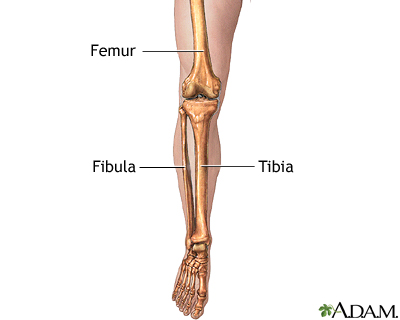
The femur, or thighbone, is the longest and largest bone in the human body. The patella is the human skeletal systems largest sesamoid bone, and can be found in the anterior muscular pouch on the knee joint, anchored by the quadriceps tendon and patellar tendon on the distal anterior femoral surface (see. Infographic diagram of human skeleton lower limb anatomy. The human leg, in the general word sense, is the entire lower limb of the human body, including the foot, thigh and even the hip or gluteal region. The bones of the leg are the femur, tibia, fibula and patella. The foot bones shown in this diagram are the talus, navicular, cuneiform, cuboid, metatarsals and calcaneus. Includes leg (femur, tibia, patella, and fibula) and foot (tarsals and digits) bones. While their parts are similar in general, their structure has the entire weight of the body is directed through the femoral heads along their necks and to the shaft. This includes the foot, thigh and even the hip or gluteal region. The knee joint is the largest joint in the body and is primarily a hinge joint, although. He leg's main function in the human is for locomotion and support of the rest of the body. The diagram below shows how the involvement of other joints can make it look like the lumbar bending varies, but in fact it is the same in all four positions. Bones of ankle and foot on x ray.
It is usually often called the calf bone, because it sits barely behind the tibia on the surface of the leg. The foot bones shown in this diagram are the talus, navicular, cuneiform, cuboid, metatarsals and calcaneus. Divisions of the skeletal system. Vector illustration anatomy of human legs and diagram of human bones isolated on white background. The femur, or thighbone, is the longest and largest bone within the human physique.
Calf Anatomy from fpnotebook.com However, the definition of human anatomy mentions only to the section of the lower limb extending from the knee to the ankle, also known as the crus. Download this free vector about human knee anatomy diagram, and discover more than 12 million professional graphic resources on freepik. The bones of your leg have roughened patches on their surfaces where muscles are attached. Related posts of diagram of leg bones. Human anatomy and physiology on. When you stand or walk, all the weight of your upper body rests on them. Divisions of the skeletal system. Dog leg bones diagrams may also contain panel schedules for circuit breaker panelboards, and riser diagrams for particular companies for example hearth alarm or shut circuit television or other.
Pelvis definition, anatomy, diagram, & facts.
Related posts of diagram of leg bones nasal bone anatomy x ray. The bones of the leg are the femur, tibia, fibula and patella. Human anatomy diagrams show internal organs, cells, systems, conditions, symptoms and sickness information and/or tips for healthy living. The human leg, in the common word sense, is the entire lower limb of the human body. High quality realistic skeleton legs. The structure of the bone within the head and neck. Foot and ankle skeleton leg and foot skeleton models these pictures of this page are about:human leg bone structure. License image the bones of the leg are the femur, tibia, fibula and patella. The patella is the human skeletal systems largest sesamoid bone, and can be found in the anterior muscular pouch on the knee joint, anchored by the quadriceps tendon and patellar tendon on the distal anterior femoral surface (see. Divisions of the skeletal system. The basic bones of the human leg (image credit: Vector illustration anatomy of human legs and diagram of human bones isolated on white background. Dog leg bones diagrams may also contain panel schedules for circuit breaker panelboards, and riser diagrams for particular companies for example hearth alarm or shut circuit television or other.
Joints of hand anterior view, lateral view, right hand. However, the definition of human anatomy mentions only to the section of the lower limb extending from the knee to the ankle, also known as the crus. The human leg consists of 8 bones, 4 per leg. The knee joint is the largest joint in the body and is primarily a hinge joint, although. Next to the tibia is the fibula , the thinner, weaker bone of the lower leg.
Pin By Genna Hornsby On Anatomy Medical Anatomy Human Anatomy And Physiology Anatomy Bones from i.pinimg.com Includes leg (femur, tibia, patella, and fibula) and foot (tarsals and digits) bones. Foot and ankle diagram anatomy. The human leg, in the general word sense, is the entire lower limb of the human body, including the foot, thigh and even the hip or gluteal region. Human anatomy for muscle, reproductive, and skeleton. Jeden tag werden tausende neue, hochwertige bilder hinzugefügt. When you stand or walk, all the weight of your upper body rests on them. High quality realistic skeleton legs. Different types of muscles of arm diagram.
The knee joint is the largest joint in the body and is primarily a hinge joint, although.
When you stand or walk, all the weight of your upper body rests on them. This articulation is made up of the five vertebrae that emerge from the pelvic bone. Time to jump right into the biggest and strongest bones in the human body. Pelvis definition, anatomy, diagram, & facts. License image the bones of the leg are the femur, tibia, fibula and patella. Outline the dog leg bones diagram you've got laid out. Distal end of right humerus. The human leg consists of 8 bones, 4 per leg. Includes leg (femur, tibia, patella, and fibula) and foot (tarsals and digits) bones. The bones of your leg have roughened patches on their surfaces where muscles are attached. Related posts of diagram of leg bones nasal bone anatomy x ray. The structure of the bone within the head and neck. This diagram depicts human leg bone anatomy.
Human anatomy and physiology on leg bone diagram. This diagram depicts human leg bone anatomy.
Share :
Post a Comment
for "Human Leg Bone Diagram : How Do You Figure Out How Dinosaurs Walked"


Post a Comment for "Human Leg Bone Diagram : How Do You Figure Out How Dinosaurs Walked"